* Welcome *

You are number...
- click the words for navigation -


| F1 Karen (Dim Sum Dolly) | M1 Lucas (EMO boy) |
| F2 Delia (Chua Hong Hong) | M2 Alvin (Guest-Of-Honor) |
| F3 Miki (pied piper) | M3 Zhao Zhi (Mr. BOO) |
| F4 Flavia (Ms. pokey) | M4 Mr. Tung Zhi Kang (Kun-jun virus) |
| F5 Siti (sweet lil thing) | |
| F6 Sajini (she smells nice) | |
| F7 Faith (she gives us hope) |


 |
 |
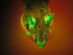 |
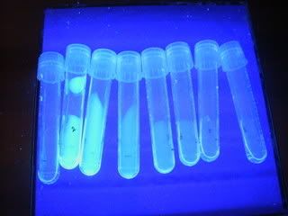
The protein that is responsible for the fluorescent glow in the mouse is called Green Fluorescent Protein, also known as GFP. in this experiment, we will be scaling up colonies of E.coli that is been inserted with GFP.
What is GFP?
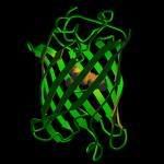
GFP is a protein that has a unique β-barrel structure that consists of 11 β-strands + 1 α-helix + chromophore running through the middle. GFP is one of the most commonly used bioreporter of a gene-expression or gene product at the molecular level.
Though GFP was first isolated and purified in the 1960s by Osamu Shimomura from Aequorea victoria, a type of jellyfish., the true potential of GFP was not comprehended until in 1987 when Douglas Prasher first thought of the idea of using GFP as a ‘tag’ that reports whenever a particular protein was being produced by a cell. Since a protein molecule was extremely small to be observed unless under an electron microscope, why not attach a marker so that it can be easily detected even by the naked eye?
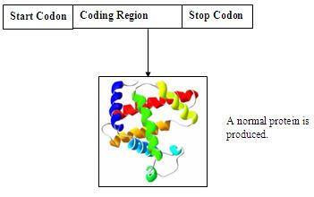
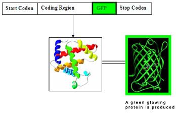
So HOW GFP is inserted into the nucleotide sequence and expressed, for example in the case of this experiment, an E.coli??
A simplify diagram of a nucleotide sequence
A simplify diagram of a GFP inserted nucleotide sequence
Overall Objectives
The main objective of the whole practical is … *drum roll*… To be able to get an A+++ for our report!!!!!! Hahaha just kidding, well that’s about 80% of the whole objective. The other 20% is to learn and experience the idea of scaling up the growth of E.coli inserted with GFP in a step-wise manner from a seed culture all the way to a bioreactor. At the end of the experiment, we also aim to be able to:
For purification, extraction that was carried out during the isolation step will be purified using Gel Permeation or Size Exclusion Chromatography. this method uses a column of polymer resins (Sephadex G75).
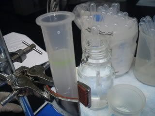
First up, we had to label eight test tubes (1 to 8) and a blank where the blank was filled with 2 ml of ammonium bicarbonate. Next is to carefully drain the column into a waste beaker until the buffer is just even with the top of the gel bed, ensuring that the column does not run dry. (water water!) we had to use a disposable plastic pipette to transfer the cell-free extract to the top of the gel by gently swirling the pipette around the inside edge of the column, just above the top of the packed matrix. (Mmmm!)